
Image Fusion for Brain Tumor Diagnosis Using Fractal Weighted Adaptive Dual Channel Neural Network
DOI:
https://doi.org/10.22401/Keywords:
Image fusion, local extrema, Enhanced Weighted Pulse, Dual Channel Neural Network, Fractal Dimension, Differential Box Counting, Brain imagingAbstract
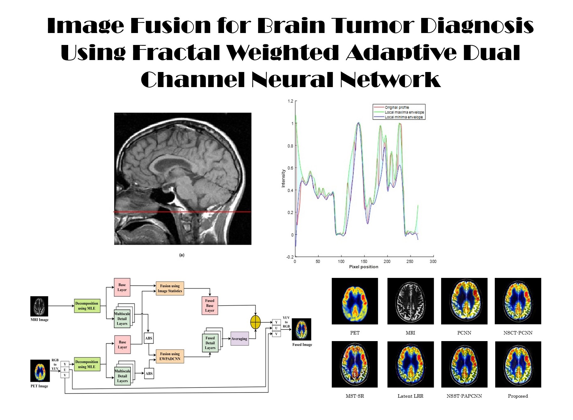
This research paper proposes a novel image fusion algorithm for assisting effective diagnosis of brain cancers. The method extracts detailed features from source images using a multi-level decomposition strategy that leverages local extrema information. The detailed layers are fused using an Enhanced Weighted Pulse Adaptive Dual Channel Neural Network (EWPADCNN), with fusion weights calculated using the Fractal Dimension with Differential Box Counting (FDDBC) method. In order to preserve low-frequency information, the base layers uses a weighted average method based on pixel significance. Comprehensive tests on 100 slices from four different datasets of brain disorders demonstrates that the proposed approach outperforms current fusion methods in both qualitative and quantitative assessments. These results support the suggested method as an efficient and trustworthy technique for improving brain imaging diagnostic quality. Quantitative evaluation demonstrates average improvements of 41%, 5%, 16%, 23%, 34%, and 4% in terms of AG, H, SF, MI, QABF, and SD metrics, respectively, compared to existing fusion methods.
References
[1] Yin, M.; Liu, X.; Liu, Y.; Chen, X.; “Medical image fusion with parameter-adaptive pulse coupled neural network in non-subsampled shearlet transform domain”. IEEE Trans. Instrum. Meas., 68 (1): 49–64, 2019.
[2] Du, J.; Li, W.; Lu, K.; Xiao, B.; “An overview of multi-modal medical image fusion”. Neurocomputing, 215: 3–20, 2016.
[3] Guo, K.; Hu, X.; Li, X.; “MMFGAN: A novel multimodal brain medical image fusion based on the improvement of generative adversarial network”. Multimedia Tools Appl., 81 (4): 5889–5927, 2022.
[4] Li, S.; Kang, X.; Fang, L.; Hu, J.; Yin, H.; “Pixel-level image fusion: A survey of the state of the art”. Inf. Fusion, 33: 100–112, 2017.
[5] Chao, Z.; Duan, X.; Jia, S.; Guo, X.; Liu, H.; Jia, F.; “Medical image fusion via discrete stationary wavelet transform and an enhanced radial basis function neural network”. Appl. Soft Comput., 118, 108542,2022.
[6] Yu, N.; Li, J.; Hua, Z.; “Decolorization algorithm based on contrast pyramid transform fusion”. Multimedia Tools Appl., 81: 15017–15039, 2022.
[7] Aishwarya, N.; Bennila Thangammal, C.; Praveena, N.G.; “NSCT and focus measure optimization based multi-focus image fusion”. J. Intell. Fuzzy Syst., 41: 903–915, 2021.
[8] Kamarthi, V.; Satyanarayana, D.; Ninjappa, G.P.M.; “Multimodal medical image fusion based on intuitionistic fuzzy sets and weighted activity measure in NSST domain”. Curr. Signal Transduct. Ther., 17: 22–31, 2022.
[9] Johnson, J.L.; Padgett, M.L.; “PCNN models and applications”. IEEE Trans. Neural Netw., 10 (3): 480–498, 1999.
[10] Vanitha, K.; Satyanarayana, D.; Prasad, M.G.; “Multi-modal medical image fusion algorithm based on spatial frequency motivated PA-PCNN in the NSST domain”. Curr. Med. Imaging, 17: 634–643, 2021.
[11] Singh, S.; Gupta, D.; Anand, R.S.; “Non-subsampled shearlet based CT and MR medical image fusion using biologically inspired spiking neural network”. Biomed. Signal Process. Control, 18: 91–101, 2015.
[12] Guo, Z.; Song, Y.; Zhao, Y.; “An adaptive infrared image segmentation method based on fusion SPCNN”. Signal Process.: Image Commun., 87: 115905, 2020.
[13] Yang, Y.; Gao, C.; Ming, Z.; Guo, J.; Leopold,E.; Cheng,J.; Zuo,J.; Zhu,M.; "LatLRR-CNN: An infrared and visible image fusion method combining latent low-rank representation and CNN". Multimedia Tools and Applications, 82 (23): 36303-36323, 2023.
[14] Liu, Y.; Liu, S.; Wang, Z.; “A general framework for image fusion based on multi-scale transform and sparse representation”. Inf. Fusion, 24: 147–164, 2015.
[15] Ibrahim, S.I.; El-Tawel, G.S.; Makhlouf, M.A.; “Brain image fusion using the parameter-adaptive pulse coupled neural network (PA-PCNN) and non-subsampled contourlet transform (NSCT)”. Multimedia Tools Appl., 83 (9): 27379–27409, 2024.
[16] Xiaomin, L.; Haowen, Y.; “An algorithm to generate a weighted network Voronoi diagram based on improved PCNN”. Appl. Sci., 12 (12): 6011, 2022.
[17] Venkata Srikanth, M.; Suneel Kumar, A.; Nagasirisha, B.; Lakshmi, T.; “Brain MRI and CT image fusion using multiscale local extrema and image statistics”. ECTI Trans. Electr. Eng. Electron. Commun., 22 (1):1-11, 2024.
[18] Panigrahy, C.; Seal, A.; Mahato, N.K.; Bhattacharjee, D.; “Differential box counting methods for estimating fractal dimension of gray-scale images: A survey”. Chaos Solitons Fractals, 126: 178–202, 2019.
[19] Chen, Y.; Park, S.K.; Ma, Y.; Ala, R.; “A new automatic parameter setting method of a simplified PCNN for image segmentation”. IEEE Trans. Neural Netw., 22 (6): 880–892, 2011.
[20] Otsu, N.; “A threshold selection method from gray-level histograms”. Automatica, 11: 23–27, 1975.
[21] Shreyamsha Kumar, B.K.; “Image fusion based on pixel significance using cross bilateral filter”.Signal Image Video Process, 9: 1193–1204, 2015.
Downloads
Published
Issue
Section
License
Copyright (c) 2026 Shailaja Mantha, Jatothu Brahmaiah Naik, Kanagala Sateesh Kumar, Rajanidevi

This work is licensed under a Creative Commons Attribution 4.0 International License.

.jpg)